Honors Human Physiology
BIOSC 1070, NROSCI 1070, MSNBIO 2070
Fall Semester 2020
BIOSC 1070, NROSCI 1070, MSNBIO 2070
Fall Semester 2020
As an introduction, watch this movie.
If the movie does not play in this window, or you would like to see it in a window of alternate size, download it from this link.
 |
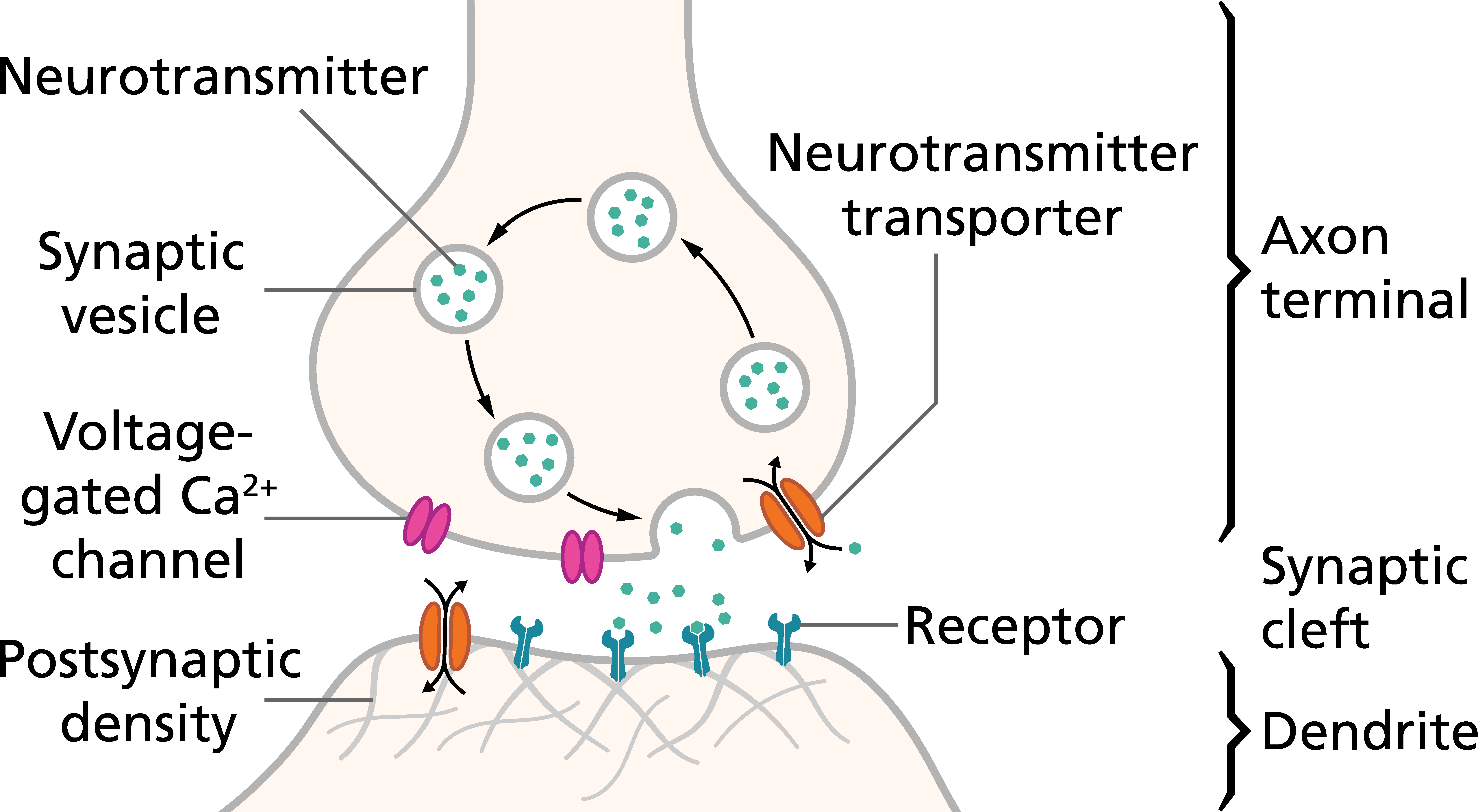
Neurotransmitters are chemicals that transmit signals from one neuron to the next. Neurotransmitters are packaged into vesicles located in the nerve terminal. When an action potential depolarizes the nerve terminal, voltage-gated Ca2+ channels are opened, allowing Ca2+ to enter the terminal. When the calcium ions enter the presynaptic terminal, they bind with special protein molecules on the inside surface of the presynaptic membrane, called release sites. This binding in turn causes the release sites to open, allowing a few transmitter vesicles to release their transmitter into the synaptic cleft (space between a nerve terminal and the dendrites or soma of another neuron). The released neurotransmitter binds to receptors on the dendrite or soma of the postsynaptic neuron. |
Let's make sure the terminology is clear:
The nature of information processing by the nervous system requires that neuronal signaling occur over a very short temporal period. When an action potential reaches the nerve terminal, the neurotransmitter that is released must have only a transient effect on the postsynaptic neuron. This is accomplished by removing the neurotransmitter from the synaptic cleft soon after it is released. There are two main mechanisms through which this is completed:
Neurotransmitters have one of two general effects when they bind to postsynaptic receptors:
There are several types of second messenger systems. One of the most common types entails the activation of G proteins. In the example below, the inactive G protein complex in the cytosol consists of guanosine diphosphate (GDP) plus three components: an alpha (α) component that is the activator portion of the G protein, and beta (β) and gamma (γ) components that are attached to the alpha component. As long as the G protein complex is bound to GDP, it remains inactive. When the receptor is activated by a neurotransmitter, the receptor undergoes a conformational change, exposing a binding site for the G protein complex, which then binds to the receptor. This causes the α subunit to release GDP and simultaneously bind guanosine triphosphate (GTP) while separating from the β and γ portions of the complex. The separated α-GTP complex is then free to move within the cytoplasm and have a number of potential effects.

Although a particular second messenger system can cause different effects in different neurons, one or more of the following effects are induced (as indicated in the diagram above):
Although neurotransmitters can elicit a number of complex changes in the postsynaptic neuron, often these effects are categorized as excitatory or inhibitory. After all, the key aspect of neuronal signaling is the generation of action potentials, so it is logical to divide neurotransmitter effects into those that:
For example, an excitatory neurotransmitter could open a ligand-gated Na+ channel and cause depolarization of the neuron, decrease conductance through K+ channels, or induce the synthesis of additional ligand-gated Na+ channels. An inhibitory neurotransmitter could open Cl- channels and cause hyperpolarization of the neuron, increase conductance through K+ channels, or induce the synthesis of additional K+ leak channels.
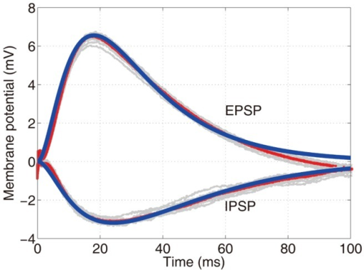
Although neurotransmitters can elicit complex postsynaptic effects, often they cause sudden changes in ionic movements across the membrane. These ionic movements produce the postsynaptic potentials that are added at the axon hillock; if the addition amounts to a sufficient depolarization, an action potential will occur.
 |
Excitatory postsynaptic potentials (EPSPs) occur when positive ions enter the neuron or negative ions leave. In most cases, the EPSP is a result of opening of cation channels that allow both Na+ and K+ to pass across the membrane. However, the conductance for Na+ usually predominates, such that a depolarization occurs in the neuron. Inhibitory postsynaptic potentials (IPSPs) occur when negative ions (such as Cl-) enter the neuron or positive ions leave. |
As an EPSP or IPSP propagates passively down a dendrite, it is altered by the physical properties of the neuronal membrane. This distorts the shape of an EPSP or an IPSP, so that if the electrical potential was recorded at a distance from the synapse where the potential was generated, it would look quite different that near the synapse. By use of cable theory, the change in the properties of an EPSP or IPSP as it moves down a membrane can be modeled mathematically. However, this is certainly beyond the scope of this course.
WHAT DO I NEED TO KNOW?
Most synapses are axo-dendritic (i.e., the synapse is on a dendrite) or axo-somatic (i.e., the synapse is on the neuronal soma), as described above. However, other synaptic configurations also exist. One such configuration is the axo-axonic synapse, where an axon of one neuron synapses on the axon of another neuron.
Although we normally think of neurotransmitters binding to postsynaptic receptors, some nerve terminals have autoreceptors for the neurotransmitter they release. Often these autoreceptors play a homeostatic role, and serve to adjust the amount of neurotransmitter released subsequently. In other words, autoreceptors serve to assure that a nerve terminal is not releasing too much, or too little, neurotransmitter.
Lets review these concepts by watching a movie from the KhanAcademy.
If the movie does not play in this window, or you would like to see it in a window of alternate size, download it from this link.
The most common neurotransmitters are amino acids, particularly glutamate, GABA (gamma-aminobutyric acid), and glycine. The actions of these amino acids will be discussed in subsequent modules. Glutamate is an excitatory neurotransmitter, and is released by most sensory afferents as well as neurons in the central nervous system. GABA and glycine are inhibitory neurotransmitters; glycine is mainly released from spinal cord neurons, whereas GABA is released from neurons in many brain areas. In fact, GABA is the most common inhibitory neurotransmitter present in the central nervous system.
Another very common neurotransmitter is acetylcholine. Acetylcholine is released by neurons in many brain regions, as well as by motoneurons, sympathetic and parasympathetic preganglionic neurons, and parasympathetic postganglionic neurons. Acetylcholine is mainly an excitatory neurotransmitter, although there are some examples where it has an inhibitory role (e.g., parasympathetic postganglionic nerve fibers that lower heart rate).
Another group of important neurotransmitters are the monoamines, chemicals that contain one amino group that is connected to an aromatic ring by a two-carbon chain (-CH2-CH2-). The monoamine neurotransmitters include dopamine, norepinephrine, and serotonin. Monoamine neurotransmitters typically bind to G-protein coupled receptors, such that they can produce a wide variety of effects in the postsynaptic neuron. Many neurons that release monoamine neurotransmitters branch extensively, such that each neuron makes synapses on a large number of postsynaptic neurons. Many neurological and psychiatric diseases are related to the monoamine neurotransmitters.
Nitric oxide (NO) is released by nerve terminals in areas of the brain responsible for long-term behavior and memory. NO is different from other small-molecule transmitters, as it is very lipophilic and cannot be stored in the nerve terminal. Instead, it is synthesized as needed and then diffuses out of the presynaptic terminals to affect adjacent neurons. NO usually does not alter the membrane potential, but instead changes intracellular metabolic functions that modify neuronal excitability for seconds, minutes, or perhaps even longer. NO is also an important signaling molecule in the cardiovascular system, as you will learn in August.
A variety of peptides called neuropeptides can also act as neurotransmitters, but they usually have very specialized functions. For example, some hypothalamic neurons release peptides that control the release of hormones from the anterior pituitary. In some cases, a peptide hormone is co-released with one of the "classical" neurotransmitters discussed above.
Lets review these concepts by watching a movie from the KhanAcademy.
If the movie does not play in this window, or you would like to see it in a window of alternate size, download it from this link.