Honors Human Physiology
BIOSC 1070, NROSCI 1070, MSNBIO 2070
Fall Semester 2020
BIOSC 1070, NROSCI 1070, MSNBIO 2070
Fall Semester 2020
As an introduction, watch this movie from the KhanAcademy.
If the movie does not play in this window, or you would like to see it in a window of alternate size, download it from this link.
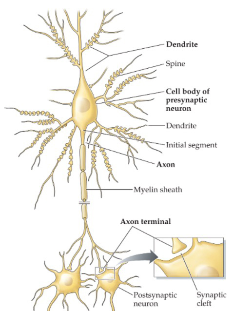
Not all neurons are the prototypical cells described in the KhanAcademy video. In general, neurons receive inputs from other nerve cells on the dendrites and cell body, and transmit outputs to other neurons or effectors like muscle cells through an axon. Most neurons have multiple dendrites and one axon, which can branch near where it reaches its target. However, there are some exceptions.
To start, let's discuss the general classes of neurons, and their anatomy:
 |
Interneurons are the most common cells in the central nervous system. The processes of interneurons always remain within the central nervous system, although some projection (or relay) interneurons may have long axons that extend for considerable distances (i.e., from the brain to the spinal cord). Most interneurons are multipolar neurons, as they have many dendrites as well as an axon that branches to multiple targets. Thus, one interneuron can receive synaptic inputs on their dendrites and cell body from thousands of neurons, and in turn send outputs to many other interneurons, or perhaps to cells that provide outflow from the nervous system to peripheral targets like muscle or glands. Such output cells are discussed below. Interneurons are integrators, and the main components of the neural circuits that process information in the nervous system. The neuron pictured on the left has dendritic spines. Dendritic spines are typically sites of excitatory synapses, where synaptic transmission makes it more likely that the neuron will generate an action potential. Inhibitory synapses are usually located on the shaft of the dendrites, the cell body, and the initial segment. Inhibitory synaptic transmission makes it less likely that a neuron will generate an action potential. |
Sensory afferent neurons carry inputs from sensors in the periphery to the central nervous system. The term "afferent" means "carrying into," and usually describes the transmission of information towards the brain and spinal cord. The dendrites of sensory afferent neurons are often specialized to receive inputs from a peripheral sensory receptor (as in the vestibular and auditory systems), or may be a component of the peripheral sensory receptor (as in the skin and muscle). Usually, the cell bodies of sensory afferent neurons are coalesced in a ganglion outside the central nervous system.
 |
Most sensory afferent neurons are pseudounipolar (neuron 1 to the left) or bipolar (neuron 2 to the left). Pseudounipolar neurons (neuron 1 to the left) have one projection from the cell body, which splits into two axons: one that extends into the periphery and one that extends into the central nervous system. Afferents that project into the spinal cord from skin and muscle are typically pseudounipolar. The cell bodies of these afferents are located in the dorsal root ganglia near the spinal cord, as will be discussed during the first week of class. The peripheral branch extends through a peripheral nerve, and is part of the sensory receptor in skin or muscle. The central branch projects through the dorsal (posterior) root into the spinal cord, and terminates on interneurons or motoneurons in the spinal cord, and may even project to the brainstem. Some afferents entering the brainstem through the cranial nerves are also pseudounipolar. Other sensory afferent neurons with projections through cranial nerves are bipolar (neuron 2 to the left). One branch from the cell body is effectively a dendrite, and extends into the periphery to make synaptic contact with sensory receptors. In other words, the sensory receptors deposit neurotransmitters onto receptors on the dendrite of the bipolar neuron. The other branch from the cell body is effectively an axon, and travels through a cranial nerve to the brainstem. Almost always, the cell bodies of bipolar neurons are in a ganglion outside the nervous system, although we will encounter one exception during the course. |
Efferent neurons send their axons out of the central nervous system, to control effectors in the periphery. These include motoneurons that innervate muscle fibers and sympathetic and parasympathetic preganglionic neurons that regulate autonomic function. We will discuss these efferent neurons later in the course.